Psychoacoustic Evaluation
Psychoacoustic evaluation is to examine voice disorder in an
objective manner.
1. GRBAS scale
It evaluates grade of hoarseness, roughness, breathy voice,
asthenic voice and strained voice on a scale of 0 to 4 grade.
2. Cons ensus Auditory Perceptual Analysis (CAPE-V)
It assesses the grade of hoarseness on a visual analog scale
of 0~100 in a subjective manner.
3. VHI(Voice Handicap Index)
Before the comprehensive voice examination, patient evaluates his/her own voice condition subjectively. When evaluating, the patient reads the given reading passage for 2~3 minutes at his/her usual voice pitch and loudness while the evaluator listens and objectively evaluates the level of hoarseness according to the assessment methods mentioned above.
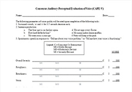 CAPE-V
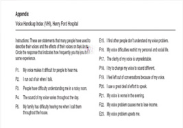 VHI
VHIPhonatory aerodynamic test
The voice is created as the air pressure from the lungs creates a vibration in the air through the larynx and passes through the pharynx to form a sound.
 Examination list
phonation time
airflow rate
air pressure
efficiency
resistance
capacity
Examination Method
Sensors are attached in all muscles related to the vocalization and respiration then MPES gets data evaluating the muscle movement, respiration and so forth in an objective way. PKE uses total 19 channels and observes movement of 12 muscles along with its respiration, vocal loudness and so forth.
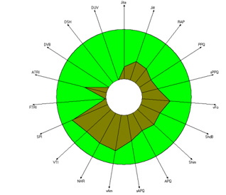 MDVP
MDVP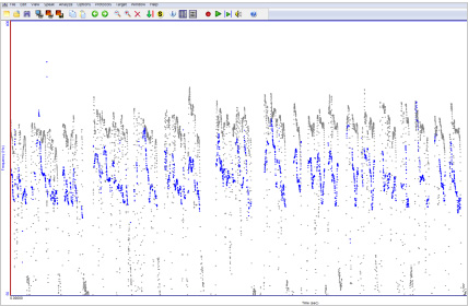 CSL
CSL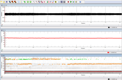 RTP
RTPVoice Range Profile Program
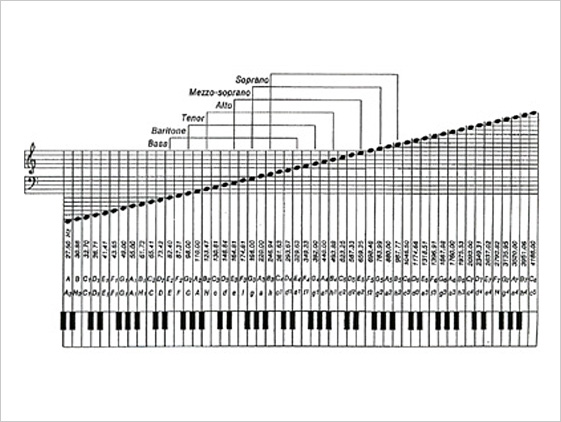
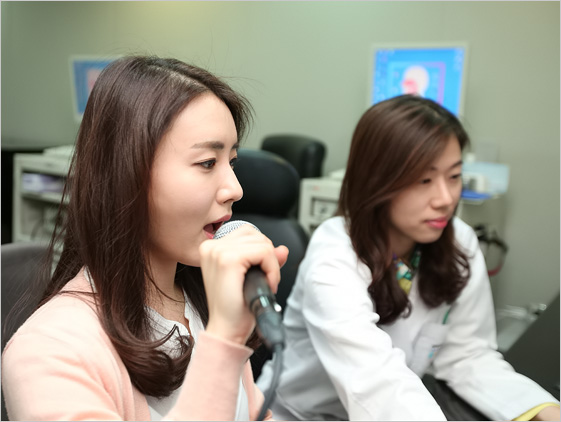
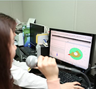
Voice range profile program shows the phonation amplitude and phonetic pattern of voice pitch range through 5 notes-scale phonation.
It checks the chest register, head register, falsetto, and voice pitch amplitude pattern. The horizontal axile shows the frequency, pitch while vertical axile shows the amplitude loudness.
Digital fiberlaryngoscopy
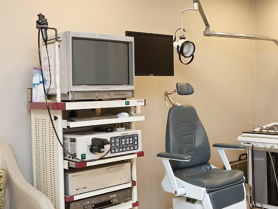
Digital fiberlaryngoscopy is used for nose, throat cavity examination, biopsy sampling, diagnostics. The fiber samples laryngoscopy with biopsy forceps, can help detect early lesions, disease of the throat cavity, after inspection is a good precision instruments.
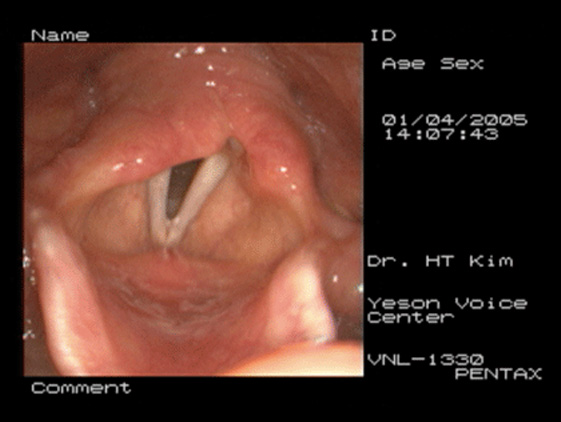
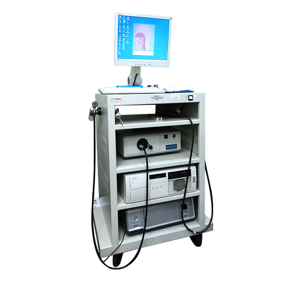
Rhino-Laryngeal Stroboscope
Rhino-laryngeal stroboscopy visualizes vocal folds movement in the video so
it enables to observe the phonatory pattern and the existence of laryngeal disease.
This is using an optical illusion of which the particular scene is remained for
0.2 seconds in the eye.
Sensors in the neck evaluates the vibratory pattern and xenon lamp reflects
vocal folds image and observe its vocal fundamental frequency and the phonatory pattern.
High-Speed Video system
High-Speed Video System captures 2000 frames per second and its strength is
that it can observe even the spasmodic dysphonia, vibration irregularity and
tension discrepancy. Also, it can also detect abnormal phonatory pattern.
Color High-Speed Video System
Color High-Speed Video System offers the image of vocal folds in vivid
color and it can capture 5000 frames per sec making it possible to observe
the real-time motion of vocal folds.
Language Evaluation
In case of a language disorder, a professional speech language and pathologist evaluates and analyzes a patient’s language ability.
In our center, the main evaluation is to assess speech disorders such as articulation disorder, dysarthria and fluency disorder (stammering).
-
 1. Articulation disorder
It poses a problem with articulation due to the structural and functional problems in that it’s hard to clearly articulate. The characteristics include omission, substitution, distortion, addition and so forth. For the evaluation, Picture Consonant Articulation Test (PCAT) and Lee-Kim Test of Korean Articulation Test and Oral Speech Mechanism Screening Examination (OSMSE-R) are held.
-
 2. Dysarthria
It’s a type of motor speech disorder. It’s a neurological damage that it induces disorders related to speech difficulty when it comes to planning, grouping, controlling neural muscles and execution. The energy required to control the respiration, resonance, vocalization, articulation when producing the word shows abnormality in power, velocity, range, strength, articulation and clarity. The muscles in the articulating organ cannot cooperate inducing articulation errors, breathy voice, uncontrollable pitch and strength disorder. Voice Examination is held to evaluate articulation problem.
-
 3. Fluency disorder
It’s a language disorder which appears in an inadequate rhythmical problem when speaking. The common problem is stammering or fast talking, neurological fluency disorder. The evaluation tool is Paradise-Fluency Assessment: P-FA which will be referred to as P-FA.
Swallowing assessment
To evaluate swallowing problems at different angles, both of visualizing and non-visualizing tools are being in use.
Each method provides partial information on the anatomy of the pharynx, swallowing or swallowing physiology.
Recently the video endoscope is being widely used to examine the structure of mouth and pharynx before or
after swallowing. This method is called FEES which stands for flexible fiberotic examination.
This evaluation can be videotaped and provides the best scenes from upside view which can observe the pharynx
structure including cover larynx, airway entrances, larynx valley, larynx cover wrinkles and so forth.
The best thing about this examination is it doesn’t get exposed to the radiation.